Agriculture
December 11, 2023
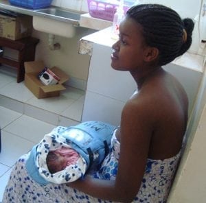
CellScope Oto
Read SolutionImplemented by
Cellscope
Updated on February 6, 2024
·Created on June 14, 2016
CellScope is a portable digital microscope that assists in the remote diagnosis of blood-borne communicable diseases.
CellScope combines optical microscopy and cellular communication to offer cell-phone microscopy. This portable digital microscope may assist in the remote diagnosis of blood-borne communicable diseases, specifically tuberculosis, malaria, and sickle cell disease.
Target SDGs
SDG 3: Good Health and Well-Being
Target Users (Target Impact Group)
Small and Medium-sized Enterprises, Public Sector Agencies, NGOs
Distributors / Implementing Organizations
Not yet available as the product is still under development for its applications to global health and disease diagnostics. The technology is being commercialized through CellScope, Inc., a for-profit spinout that was founded in 2010. CellScope, Inc.'s first product is an iPhone otoscope that builds upon CellScope's technology and enables parents and physicians to remotely diagnose ear infections in children, which is now commercially available.
Competitive Landscape
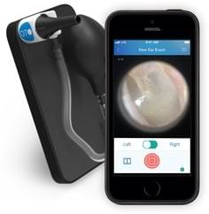
Direct competitors include CellScope Oto and Foldscope.
Regions
West Africa
Countries
Cameroon, Congo (Brazzaville), India, Thailand, Vietnam
Manufacturing/Building Method
Information not available, as the CellScope technology for global health applications is still under research and development and not yet commercially available.
Intellectural Property Type
Patent
User Provision Model
The product is not yet commercially available.
Distributions to Date Status
None, as CellScope and its applications to disease diagnostics are still under research and development.
Illumination source
Ambient light (without a condenser), white light-emitting diode (LED) for illumination in darker conditions, color LED for fluorescence imaging
Magnification level
8-120X
Spatial Resolution
1.2 micrometers
Diagnostic Applications
Tuberculosis, malaria, and sickle cell disease
Power supply type
Recharging only (smart phone powers device)
Consumables
Microscope slides
Indispensable equipment for function (Y/N)
Y
Maintenance or calibration required by user at time of use? (Y/N)
Y
Design Specifications
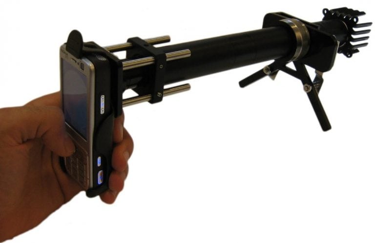
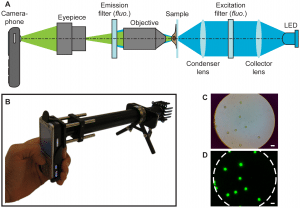
The CellScope is comprised of a mobile phone outfitted with standard microscopy optics parts (e.g. an eyepiece, tube, lenses, and light source). The functional prototypes consisted of the Nokia N73 camera phones, equipped with a 3.2 megapixel (2048×1536 pixel) CMOS camera with a 5.6×4.2 mm sensor, yielding an ?2.7 µm pixel spacing. Similar off-the-shelf mobile phones with sufficient camera systems can be leveraged. The phone and optical components were mounted using an optical rail system, and laid out as in Figure 1a shown in the product schematics. A functional, handheld prototype is shown in Figure 1b.
The CellScope apparatus can be used for bright-field and fluorescence imaging (with additional filters and LED) of medical samples, followed by capture, analysis, and transmission of images for diagnosis. Ambient light (without a condenser) is typically sufficient for brightfield imaging, a white LED can be used for illumination in darker conditions. For fluorescence microscopy, set-up requires trans-illumination geometry incorporating an LED excitation source and filters in the optical train (see Figure 1a in the product schematics).
Brightfield images can be captured using the phone's default camera settings, with the flash disabled. Fluorescent images can be captured in the cameras “Night” mode, with the flash disabled.
Spatial Resolution: 1.2 micrometers Magnification of 5x–60x.
Product Schematics
Technical Support
None available, as product still not commercially available. Main technical support and development conducted through Fletcher Lab at UC Berkeley and its collaborators.
Replacement Components
None, as product is still not commercially available.
Lifecycle
Unknown
Manufacturer Specified Performance Parameters
Capabilities such as: bright-field and fluorescent visualization of medical samples, followed by capture, analysis, and transmission of images critical for diagnosis. Resolution high enough to diagnose malaria from blood samples and tuberculosis from sputum samples.
Vetted Performance Status
Product is still under testing and evaluation. However, studies such as the one discussed in this article, a research paper titled Accuracy of Mobile Phone and Handheld Light Microscopy for the Diagnosis of Schistosomiasis and Intestinal Protozoa Infections in Côte d'Ivoire, are evaluating the performance and effectiveness of the CellScope technology.
Safety
Users must take appropriate precautions when using electrical devices.
Complementary Technical Systems
3D printers for physical product.
Academic Research and References
Kamgno, J., Pion, S., Chesnais, C., Bakalar, M., et al., 2017, A Test-and-Not-Treat Strategy for Onchocerciasis in Loa loa–Endemic Areas. N Engl J Med, Vol. 377, pp.2044-2052
Coulibaly, J., Ouattora, M., D’Ambrosio, M., et al., 2016, Accuracy of Mobile Phone and Handheld Light Microscopy for the Diagnosis of Schistosomiasis and Intestinal Protozoa Infections in Côte d’Ivoire. PLoS Negl Trop Dis, Vol. 10
Chaisson, L., Reber, H., Phan, N., Switz, L., Nilsson, F., 2015, Evaluation of mobile digital light-emitting diode fluorescence microscopy in Hanoi, Viet Nam. Int J Tuberc Lung Dis, Vol.19, pp.1068-1072
Straus, Tamara. “A Device That Could Change Healthcare.” Blum Center, 2014.
Editors, Medgadget. “CellScope, a ‘Mobile Phone Based Clinical Microscopy for Global Health Applications’.” Medgadget, 2009.
Akst, jef. “Mobile Microscopes.” The Scientist Magazine®, 2013.
UHN_News. “Mobile, Phone-Based Microscopes Work Well in the Field with Minimal Training.” EurekAlert!, 2016.
Compliance with regulations
Product still under research and development, not available.
Evaluation methods
Clinical and field studies, along with further research and development in academic labs.
Other Information
None
Agriculture
December 11, 2023
Implemented by
Cellscope
Agriculture
December 31, 2023
Implemented by
Potential Energy

Agriculture
December 14, 2023
Implemented by
AFRIpads
Agriculture
December 2, 2024
Implemented by
BioLite
Agriculture
February 29, 2024
Implemented by
BRAC University
Agriculture
January 11, 2024
Implemented by
Caminos de Agua
Agriculture
January 17, 2024
Implemented by
Aquagenx
Agriculture
February 14, 2024
Implemented by
Diagnostics for all
Agriculture
November 10, 2023
Implemented by
Embrace Innovations
Agriculture
February 15, 2024
Implemented by
HemoCue
Have thoughts on how we can improve?
Give Us Feedback